
Brain Anatomy Every Chiropractic Student Should Know for Part 1 Boards
If you’re preparing for the NBCE exam, understanding brain anatomy is essential—not only to pass your boards, but to lay a strong foundation for clinical reasoning. The brain plays a key role in coordinating the nervous system and relaying information throughout the body, making it critical knowledge for every future chiropractor.
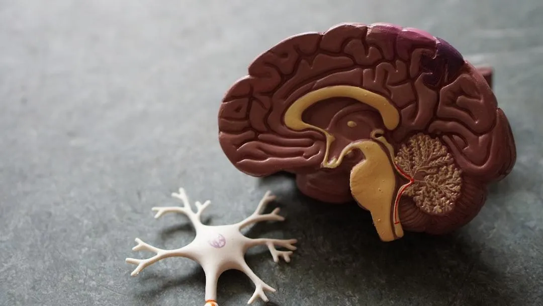
Here’s a breakdown of the most important brain structures and their functions, focusing on what you really need to know.
Cerebrum: The Brain’s Command Center
The cerebrum is the largest part of the brain, accounting for around 85% of its mass. It's divided into two hemispheres—left and right—connected by the corpus callosum, which allows communication between the two sides.
Each hemisphere contains four distinct lobes:
1. Frontal Lobe
Function: Voluntary movement, decision-making, planning, and personality.
Key Area: Broca’s Area (in the dominant hemisphere) – responsible for speech production.
Clinical Tip: Damage to this lobe can lead to motor weakness and expressive aphasia (difficulty speaking).
2. Parietal Lobe
Function: Sensory perception and spatial orientation.
Role in Practice: Integrates sensory input (touch, temperature, pain).
Clinical Tip: A lesion here may result in sensory deficits or neglect of one side of the body (especially in strokes).
3. Temporal Lobe
Function: Auditory processing, language comprehension, memory.
Key Area: Wernicke’s Area – responsible for understanding spoken language.
Clinical Tip: Damage can cause receptive aphasia (fluent but nonsensical speech), and issues with memory recall.
4. Occipital Lobe
Function: Vision.
Clinical Tip: Injury here may result in visual field deficits or cortical blindness.
Corpus Callosum: The Bridge Between Hemispheres
This thick band of nerve fibers facilitates communication between the brain’s hemispheres. It’s essential for integrating functions like motor coordination and sensory input.
Clinical Tip: In cases of severe epilepsy, the corpus callosum may be surgically severed, resulting in “split-brain” symptoms, where the two hemispheres function independently.
Basal Ganglia: Movement Modulators
The basal ganglia are deep brain structures involved in controlling and refining movement, motor learning, and behavior.
Includes: Caudate nucleus, putamen, globus pallidus.
Clinical Tip: Disorders like Parkinson’s disease originate here, causing tremors, rigidity, and bradykinesia (slowness of movement).
Bonus: Cerebellum and Brainstem
Although technically not part of the cerebrum, don’t overlook these for the NBCE Part 1 Exam:
Cerebellum
Coordinates balance, posture, and fine-tuned movement. Damage can result in ataxia (loss of coordination).
Brainstem (Midbrain, Pons, Medulla)
Controls vital functions such as breathing, heart rate, and reflexes. Damage can be life-threatening.
Final Thoughts
A clear grasp of brain anatomy helps chiropractic students understand how the nervous system influences musculoskeletal health. By focusing on the core structures—the cerebrum, lobes, basal ganglia, and related systems—you’ll be well-equipped for the NBCE Part 1 Exam and for your future clinical practice.
Pro tip: When studying, quiz yourself on which lobe or structure would be affected based on different symptoms or deficits. This helps solidify your understanding and makes exam questions feel like second nature.
Need More Help Preparing for Boards?
Copyrights 2025 | Chiro Boards Pro™ | Terms & Conditions