
Cranial Nerves: What to Know for Your NBCE Part 1 Exam
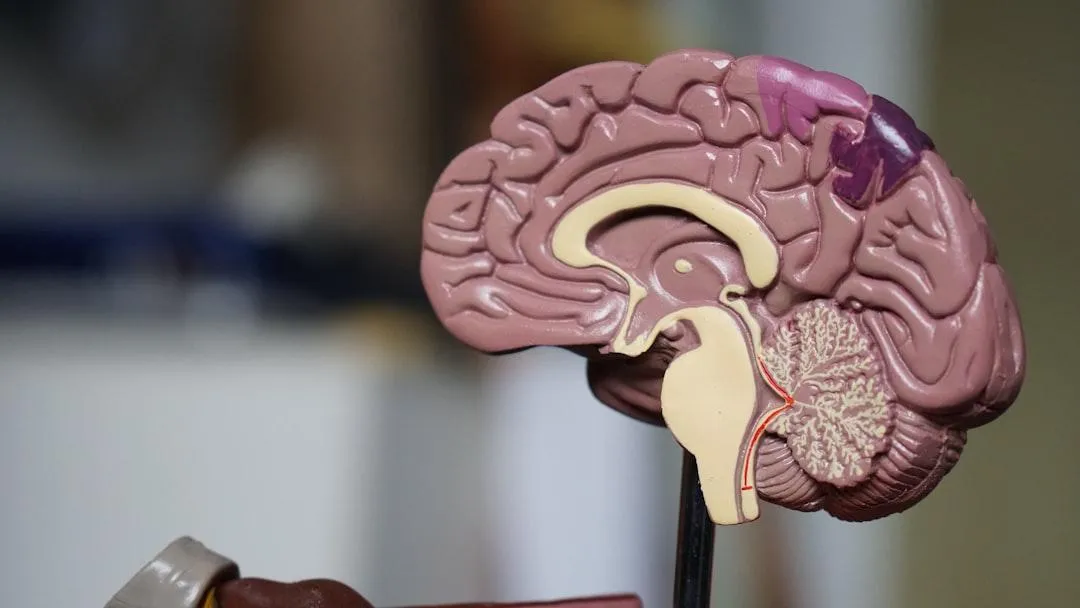
As chiropractic students, mastering the intricacies of the cranial nerves is essential for effective neurological assessments and comprehensive patient care. These twelve pairs of nerves arise directly from the brain and brainstem, each with specialized sensory, motor, or mixed roles. Below, you'll find an in-depth breakdown of each nerve’s location, function, and embryological origin — with a few helpful emojis for visual memory cues!
👃 Cranial Nerve I – Olfactory Nerve
Location: Arises from the olfactory bulb, located on the inferior surface of the frontal lobe.
Function: Sensory nerve responsible for the sense of smell.
Embryological Origin: Derived from the olfactory placode, a thickening of ectoderm in the telencephalon (part of the forebrain).
👁️ Cranial Nerve II – Optic Nerve
Location: Originates from the retina, passes through the optic canal, and forms the optic chiasm in the diencephalon.
Function: Sensory nerve transmitting visual information from the retina to the brain.
Embryological Origin: Outpouching of the diencephalon (optic vesicle).
👁️ Cranial Nerve III – Oculomotor Nerve
Location: Emerges from the anterior surface of the midbrain (mesencephalon), near the interpeduncular fossa.
Function: Motor nerve controlling most extraocular muscles (except lateral rectus and superior oblique), pupil constriction, and eyelid elevation.
Embryological Origin: Arises from the basal plate of the mesencephalon.
👁️ Cranial Nerve IV – Trochlear Nerve
Location: The only cranial nerve emerging dorsally from the brainstem, just below the inferior colliculus in the midbrain.
Function: Motor nerve for the superior oblique muscle, enabling downward and lateral eye movement.
Embryological Origin: Basal plate of the mesencephalon.
🙂 Cranial Nerve V – Trigeminal Nerve
Location: Emerges from the lateral aspect of the pons and divides into three branches: ophthalmic (V1), maxillary (V2), and mandibular (V3).
Function: Mixed nerve; sensory innervation to the face, scalp, teeth, and anterior tongue. Motor innervation to muscles of mastication.
Embryological Origin: Sensory components from neural crest cells; motor from basal plate of the metencephalon (pons).
👁️ Cranial Nerve VI – Abducens Nerve
Location: Emerges from the pontomedullary junction near the midline.
Function: Motor nerve that innervates the lateral rectus muscle, allowing the eye to move laterally (abduction).
Embryological Origin: Arises from the basal plate of the metencephalon.
🙂 Cranial Nerve VII – Facial Nerve
Location: Emerges from the pons, enters the internal acoustic meatus, and exits via the stylomastoid foramen.
Function: Mixed nerve; controls muscles of facial expression, taste from the anterior two-thirds of the tongue, and parasympathetic supply to lacrimal and salivary glands.
Embryological Origin: Motor fibers from basal plate of the metencephalon; sensory from neural crest.
👂 Cranial Nerve VIII – Vestibulocochlear Nerve
Location: Emerges at the cerebellopontine angle, enters the internal acoustic meatus.
Function: Sensory nerve responsible for hearing (cochlear division) and balance (vestibular division).
Embryological Origin: Derived from the otic placode and neural crest cells.
👅 Cranial Nerve IX – Glossopharyngeal Nerve
Location: Emerges from the lateral medulla and exits through the jugular foramen.
Function: Mixed nerve; sensory for taste in the posterior third of the tongue, monitors carotid body and sinus, motor to stylopharyngeus muscle, and parasympathetic to parotid gland.
Embryological Origin: Sensory components from neural crest; motor from basal plate of the myelencephalon.
❤️ Cranial Nerve X – Vagus Nerve
Location: Emerges from the lateral medulla, exits the skull through the jugular foramen.
Function: Mixed nerve; provides parasympathetic innervation to thoracic and abdominal organs, motor control of pharyngeal/laryngeal muscles, and sensory from external ear and viscera.
Embryological Origin: Motor components from basal plate of the myelencephalon; sensory from neural crest.
💪 Cranial Nerve XI – Accessory Nerve
Location: Has cranial and spinal roots; the spinal portion ascends through the foramen magnum and exits via the jugular foramen.
Function: Motor nerve innervating the sternocleidomastoid and trapezius muscles.
Embryological Origin: Arises from the cervical spinal cord (spinal root) and basal plate of the myelencephalon.
👅 Cranial Nerve XII – Hypoglossal Nerve
Location: Emerges from the medulla between the pyramid and olive, exits through the hypoglossal canal.
Function: Purely motor nerve; innervates intrinsic and extrinsic muscles of the tongue (except palatoglossus).Controls tongue movement for speech, food manipulation, and swallowing.
Embryological Origin: Derived from the basal plate of the myelencephalon (medulla), specifically associated with somatic efferent fibers.
Understanding the cranial nerves not only reinforces neuroanatomy but equips chiropractic students with essential tools to identify and interpret cranial nerve dysfunctions. Bookmark this guide for quick review before clinical assessments or board exams!
Need More Help Preparing for Boards?
Copyrights 2025 | Chiro Boards Pro™ | Terms & Conditions